DNA Binding Motif
| Accessions: | 1lfu_P (3D-footprint 20231221) |
| Names: | homeobox protein PBX1 |
| Organisms: | Mus musculus |
| Libraries: | 3D-footprint 20231221 1 1 Contreras-Moreira B. 3D-footprint: a database for the structural analysis of protein-DNA complexes. Nucleic acids research 38:D91-7 (2010). [Pubmed] |
| Description: | NMR Solution Structure of the Extended PBX Homeodomain Bound to DNA |
| Length: | 6 |
| Consensus: | aaTnAT |
| Weblogo: | 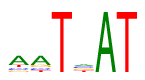 |
| PSSM: | P0 A C G T 01 54 13 13 16 a 02 54 13 16 13 a 03 0 0 0 96 T 04 24 24 24 24 n 05 96 0 0 0 A 06 0 0 0 96 T |
| Binding TFs: | 1lfu_P (Homeobox domain, Homeobox KN domain, Homeodomain leucine-zipper encoding, Homez) |
| Binding Sites: | 1lfu_A 1lfu_B |
| Publications: | Sprules T, Green N, Featherstone M, Gehring K. Lock and key binding of the HOX YPWM peptide to the PBX homeodomain. The Journal of biological chemistry 278:1053-8 (2003). [Pubmed] |
Disclaimer and license
These data are available AS IS and at your own risk. The EEAD/CSIC do not give any representation or warranty nor assume any liability or responsibility for the data nor the results posted (whether as to their accuracy, completeness, quality or otherwise). Access to these data is available free of charge for ordinary use in the course of research. Downloaded data have CC-BY-NC-SA license. FootprintDB is also available at RSAT::Plants, part of the INB/ELIXIR-ES resources portfolio.